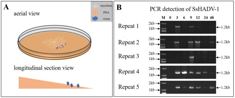
Figure 1.
(A) Schematic diagram of SsHADV-1 virion inoculation on an angled plate. White, sandy brown, and blue colors represent the mycelium, PDA, and SsHADV-1 virions, respectively. After the mycelium grew on cellophane-covered PDA for 36 h, the virions were added to the lower mycelial tips. Subsequently, the upper mycelia were collected at different time points. Briefly, 15 µL of SsHADV-1 virion (2 mg/mL) were added, and strain Ep-1PNA367 was cultured under 20 °C conditions. (B) SsHADV-1 detection in healthy tissue after mycelia inoculation with virions at different time points (0, 3, 6, 9, 12, 24, and 48 h). SsHADV-1 was detected with specific primers (Table S6) for five biological replicates. The PCR production size was about 1.2 kb.

