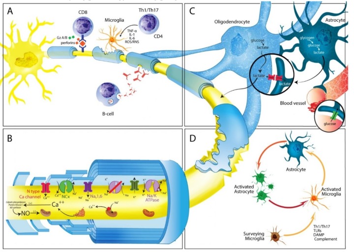
Figure 2.
Possible mechanisms involved in MS progression. (A) In progressive MS the inflammatory phenomena eventually leading to axonal degeneration and loss are compartmentalized within the CNS. Cellular components are represented by cells that come from the periphery (T and B lymphocytes), as well as by resident CNS cells (microglia cells and astrocytes). B cells can form ectopic follicle-like structures resembling tertiary lymph nodes, producing antibodies against myelin and non-myelin antigens, shown to play an important role in axonal and neuronal damage through complement cascade activation. In turn, CD8+ lymphocytes can recognize specific axonal antigens and produce tissue damage through secretion of perforin or granzymes A and B. Autoreactive CD4+ Th1 and Th17 lymphocytes can activate microglial cells, which in turn produce pro-inflammatory cytokines (IL-1, IL-6, TNF-α) or oxygen or nitrogen free radicals (ROS/RNS) causing axonal damage and neuronal loss through a bystander mechanism. (B) Following demyelination, energy requirements increase due to disruption of paranodal myelin loops. Reduction in neuronal ATP production may lead to failure of the Na+/K+ pump failure, generating a sustained sodium current, which drives reverse sodium/calcium exchange and accumulation of intra-axonal calcium. This, in turn activates degradative enzymes, including proteases, phospholipases, and calpains, resulting in further neuronal and/or axonal damage as well as impaired ATP production. (C) Axonal damage could be cause by poor trophic support. Oligodendrocytes capture glucose from circulation, breaking it down glucose to form pyruvate or lactate, which can enter axons, and be imported by mitochondria for ATP synthesis. An alternative source of energy for axons comes from glycogen stored in astrocytes, which can be transformed into glucose and later into pyruvate or lactate, depending on oxygen availability. (D) Several mechanisms cause surveillance microglia activation including Th1 or Th17 T cells; presence of microbial pathogens (PAMPs) recognized by Toll-like receptors (TLRs) or leucin-rich repeat containing receptors (NLRs); release of intracellular components from necrotic or apoptotic cells; presence of heat shock proteins, misfolded proteins (DAMPs), or components of the complement cascade. Once activated they in induce activation and proliferation of astrocytes, leading to astrogliosis.

