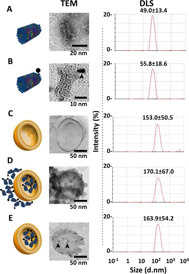
Figure 3.

Characterization of liposomes, STDO, and LSTDO. (A) STDO was synthesized and characterized by TEM and DLS. (B) To visualize STDO inside liposome, a Cd–Se quantum dot (QD, arrowhead) was conjugated to STDO. (C) Pegylated liposomes were synthesized and (D) loaded with STDO (LSTDO). DLS was able to detect only liposomes (LSTDO). (E) Excess of STDO was eliminated incubating LSTDO with a cationic resin that binds unloaded STDO but not liposomes. Arrowheads indicate QD-STDO inside liposomes.
