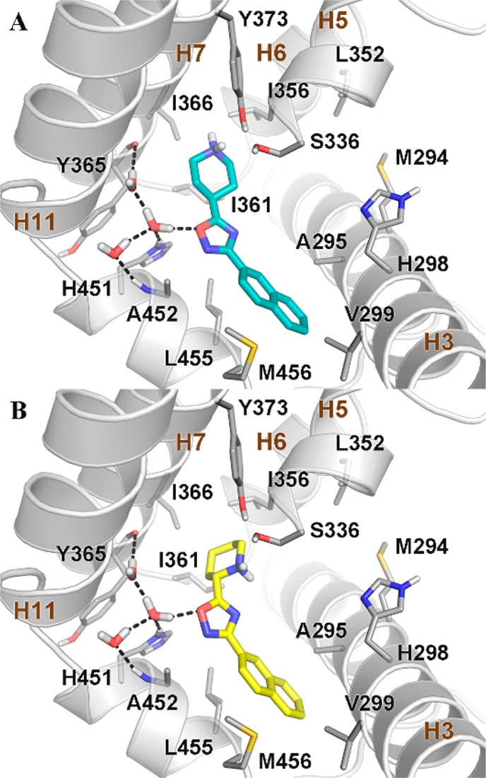
Figure 4.

Docking pose of (A) 3f (cyan sticks) and (B) 13 (yellow sticks) in the crystal structure of the FXR-LBD (PDB code: 4OIV).21 FXR is shown as gray cartoons, with amino acids important for ligand binding depicted as sticks. Nonpolar hydrogens are omitted for clarity. Hydrogen bonds are shown as dashed black lines.
