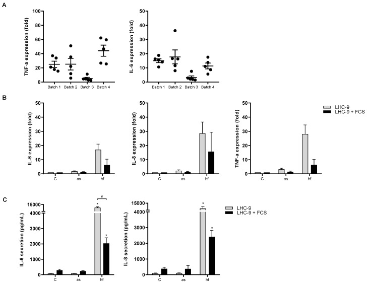
Figure 2.
Batch variations and effects of serum on cytokine/chemokine expression and release. (A) BEAS-2B cells were exposed to 100 µg dry weight/mL of four different batches of X-ray treated P. chrysogenum hyphal fragments (hf) for 6 h. The mRNA level of TNF-α and IL-6 were assessed by real time RT-PCR. Medium blanks were included as control. Plots represent mean ± SEM of 5 independent experiments. (B) BEAS-2B cells grown in LHC-9 media ± FCS were exposed to X-ray treated aerosolized spores (as) and hyphal fragments (hf) 100 µg dry weight/mL from P. chrysogenum for 6 h and assessed for expression with real-time RT-PCR or (C) cytokine/chemokine release analyzed by ELISA. Medium blanks were included as control. The results represent mean ± SEM of two or three independent experiments respectively. Statistical analyses were performed by two-way ANOVA with Dunnett’s/Sidak post-tests on log transformed data. Significant difference denoted by * (individual control vs exposed) or # (±FCS), p < 0.05.

