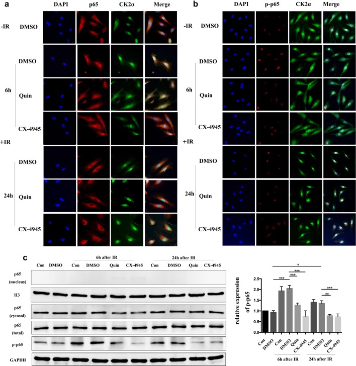
Fig. 4.
CK2 inhibition decreased IR induced phosphorylated p65 expression in HUVECs. HUVECs were pretreated with complete medium, DMSO, 25 μM Quinalizarin or 10 μM CX-4945 for 6 h, then exposed to 4 Gy IR. Cells were fixed 6 h or 24 h after IR and incubated with primary antibodies. a p65 and b p-p65 was labeled by Cy3-conjugated secondary antibody and CK2 α was labeled by FITC-conjugated secondary antibody. Cell nuclei were stained with DAPI. Fluorescence images were taken at ×400 magnification by a scanning confocal microscope. c Protein expressions of nuclear and cytoplasmic p65, total p65 and p-p65 in HUVECs, which were treated as described above, were assessed by Western blot. Mean ± SD were calculated for three independent experiments (*p < 0.05, **p < 0.01, ***p < 0.001)

