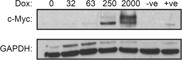
Fig. 2.

Western blot of c-Myc knock-out cells (HO15.19) stably expressing the TRE3G-cMyc transfer vector. Cells were plated at 2.0 × 10⁵ cells/ml in various dox concentrations with 2.0 ml per well in a 6-well plate. Negative (−ve) control (non-modified HO15.19), and positive (+ve) control (wild type TGR-1) cells were included. Cells were cultured for 24 h to allow for dox induced c-Myc expression, then cellular proteins were analysed by western blot using anti-c-Myc and anti-GAPDH antibodies
