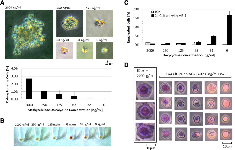
Fig. 7.
IPE cell colony forming characterization and developmental potential. a. (TOP) Colony forming assays of IPE cells at various concentrations of dox. Images of representative colonies formed after 1 week of culture in various dox concentrations. (BOTTOM) The percentage of cells that formed colonies for each concentration of dox after 7 days. b. IPE cell pellets imaged after 2 days of culture on TCP in IPE media with various dox concentrations. c. Average enucleated non-granular cell percentage after 4 days for triplicate differentiation cultures either on TCP or on a confluent layer of mouse stromal cells (MS-5). d. IPE cells and differentiated IPE cells stained using the Giemsa histology stain to allow for the identification of the cytoplasm and nuclear material. IPE cells were differentiated by co-culture on MS-5 for 4 and 8 days with 50% media exchange every 2 days. All images were taken at 400× magnification and scaled equivalently to show size variation between developmental stages

