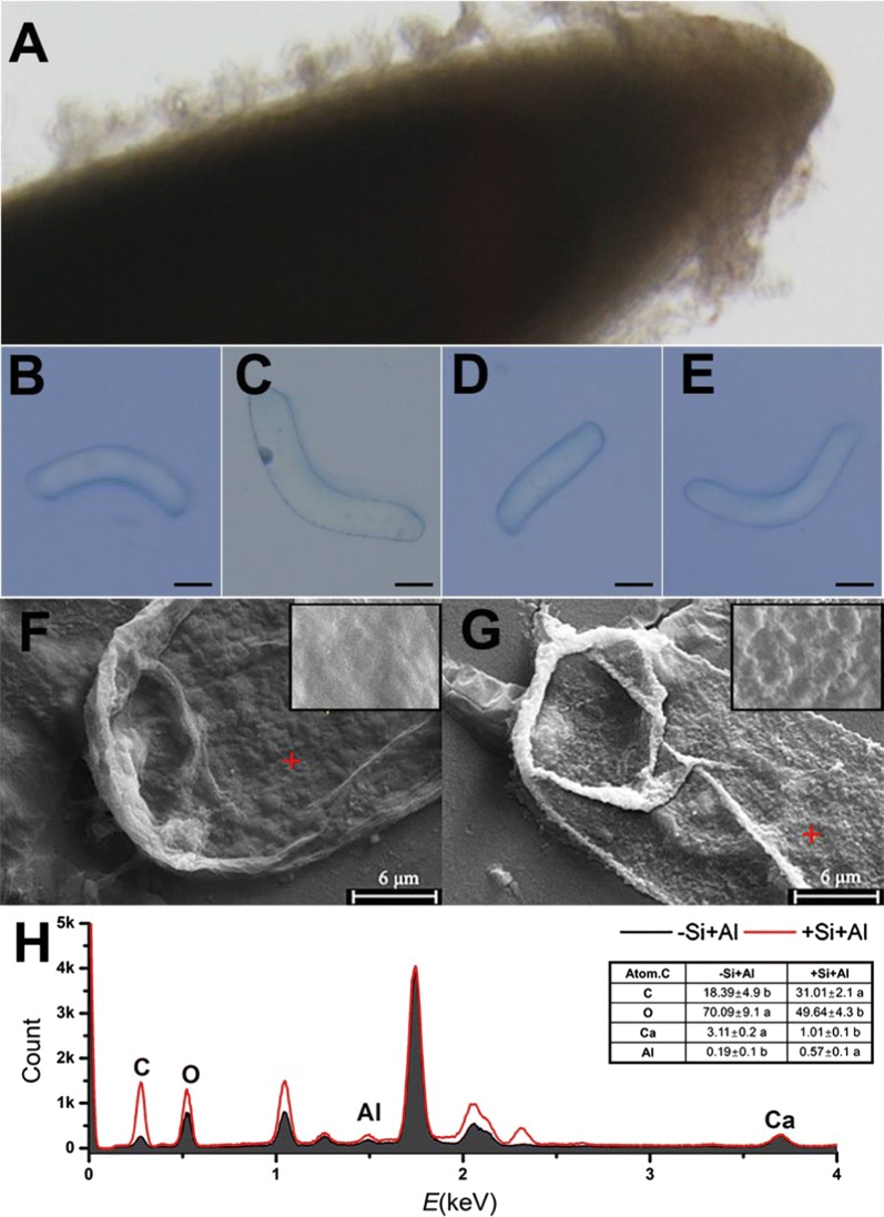
Fig. 1.
Properties of RBCs and scanning electron microscopy (SEM) image of the cells with or without the application of LBL self-assembly technique. RBCs are distributed around the root tip (A) and observed by trypan blue test. B Image of alive bare cell (−Si−Al); C Image of Al-induced dead bare cell exposed to 100 μM AlCl3 solution for 1 h (−Si+Al); D Image of silica-coat cell (+Si−Al); E Image of silica-coat cell exposed to 100 μM AlCl3 solution for 1 h (+Si+Al). Scale bars = 25 μm. RBCs was treated in 100 µM AlCl3 solution for 1 h, and then the specimens were dehydrated in ethanol and isoamyl acetate. SEM was performed for the surface images and the distribution of Ca and Al by energy dispersive spectroscopy. F Bare cells, G cells with silica-coat. Bar = 6 μm. Energy dispersive spectrometer (EDS) taken from the position of the red cross (H). The atomic content of carbon (C), oxygen (O), calcium (Ca) and aluminum (Al) was calculated

