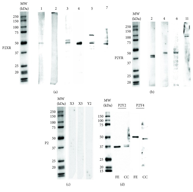
Figure 4.
Detection of P2 receptors in the clonal culture of the hOE. Cloned cells were scraped with a RIPA buffer, and cell lysates were assayed by Western blot to immunodetect P2 receptor subtypes. Representative chemiluminescent bands corresponding to P2X receptors are shown in (a) and to P2Y receptors in (b). (c) shows antibody specificity controls assessed by omission of primary antibody (P2X3) or by preadsorption of anti-P2X5 and anti-P2Y2 with the corresponding blocking peptides. The molecular weights of sample bands were corroborated using biotinylated molecular weight standards. (d) shows immunodetection of P2Y2 and P2Y4 receptors in both the fresh exfoliate (FE) sample and the clonal culture (CC) from the hOE.

