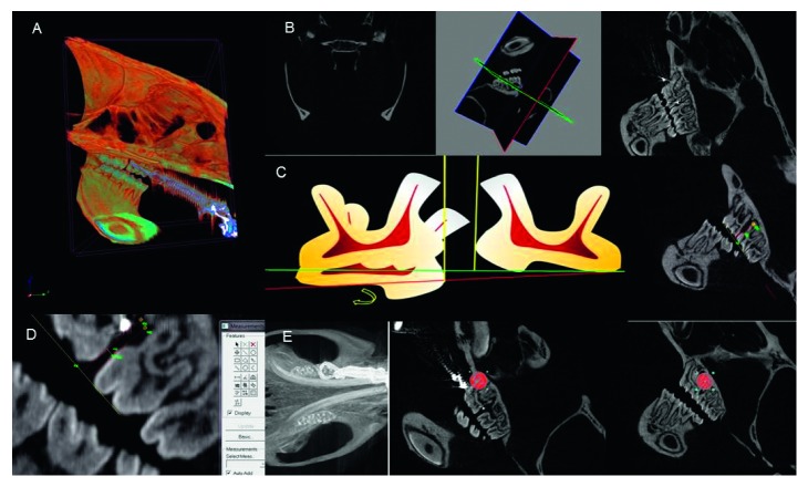
Figure 1.
(a) Acquisition of Micro-CT image. (b) Reconstructed image selected using the x, y, and z system of coordinates. (c) Measuring guides: first, schema representing the extrusion and inclination of the first molars, whose measurement guide minimizes calibration errors between the molars; second, reconstructed image chosen with the measuring guides. (d) Measurement of tooth movement through reconstructed images. (e) Reconstructed Micro-CT image showing the circular area of 60 × 60 pixels in the mesial and distal roots of the left upper first molars for measurement of bone density in the delimited region.

