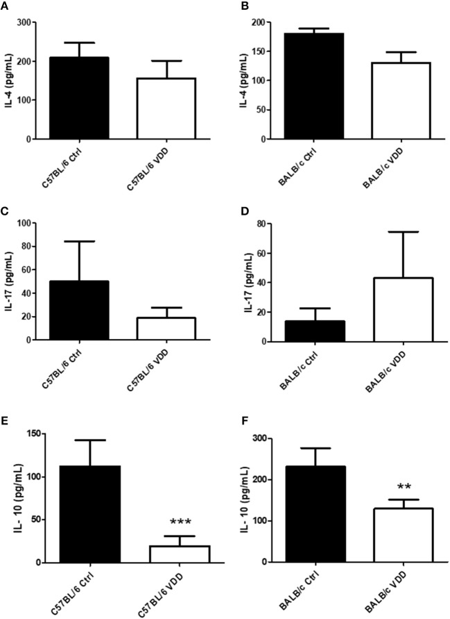
Figure 3.
Cytokine profile in the lesions of L. (L.) amazonensis-infected VDD mice. C57BL/6 and BALB/c mice normally fed (Ctrl) or on a Vitamin D-deficient diet (VDD) were subcutaneously infected in the footpad with 2 × 10⁵ L. (L.) amazonensis promastigotes. On day 92 (C57BL/6) or 99 (BALB/c) post-infection, the cytokine profile in the lesions was evaluated by ELISA. IL-4 (A,B), IL-17 (C,D), and IL-10 (E,F) levels in tissue homogenates were quantified. The data (means ± SD; n = 5; ***P < 0.001, **P < 0.01) are representative of two independent experiments producing the same result profile.

