Figure 15.

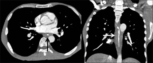
Corrosive injury of the esophagus: computed tomography (CT): axial and coronal reformatted CT images show smooth circumferential wall thickening of the mid and distal esophagus (arrows)

Corrosive injury of the esophagus: computed tomography (CT): axial and coronal reformatted CT images show smooth circumferential wall thickening of the mid and distal esophagus (arrows)