Figure 18.

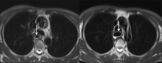
Corrosive injury of the esophagus: magnetic resonance imaging: axial T2-weighted images show mild narrowing in the mid-thoracic esophagus (left arrow) with upstream dilatation (right arrow)

Corrosive injury of the esophagus: magnetic resonance imaging: axial T2-weighted images show mild narrowing in the mid-thoracic esophagus (left arrow) with upstream dilatation (right arrow)