Figure 2.

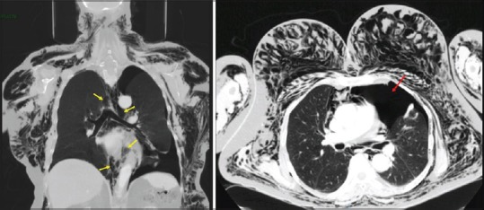
Chest computed tomography scans showing subcutaneous emphysema, pneumomediastinum (yellow arrows), and pneumothorax on the left side (red arrow)

Chest computed tomography scans showing subcutaneous emphysema, pneumomediastinum (yellow arrows), and pneumothorax on the left side (red arrow)