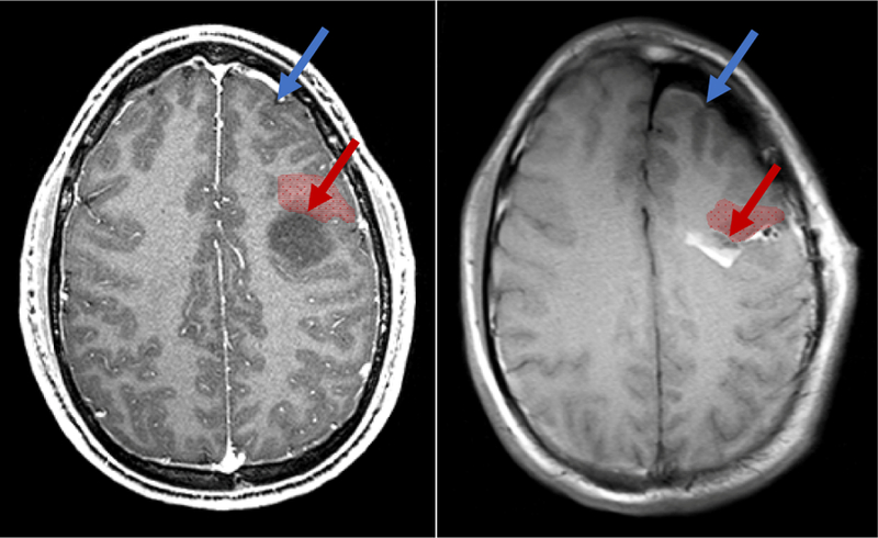
Figure 1.

A. Preoperative MRI showing a low grade glioma (dark region). B. iMRI after near-complete resection with significant brain shift. Brain shift significantly reduces the validity of neuronavigation from preoperative data. Brain shift can occur far from the surgical site (in blue). More clinically relevent, it can cause significant deformation and displacement near margins of the resection cavity (in red), precisely where surgeons could most benefit from accuracte image guidance.
