
Figure 1.
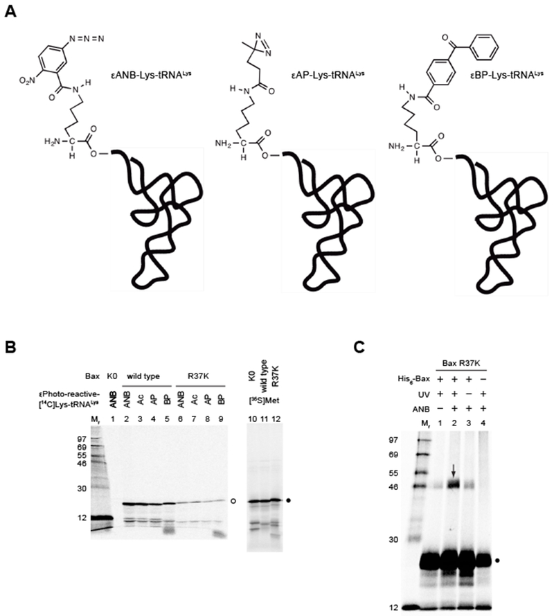
Synthesis and crosslinking of photo-reactive Bax protein. (A) Structures of photo-reactive εANB-[14C]Lys-tRNALys, εAP-[14C]Lys-tRNALys and εBP-[14C]Lys-tRNALys. (B) Synthesis of photo-reactive Bax protein. The wild type and Lys-null (K0) or single-Lys (R37K) mutant proteins are synthesized using wheat germ extract-based in vitro translation system in the presence of photo-reactive probe ANB, AP or BP-labeled [14C]Lys-tRNALys, or the acetylated (Ac) [14C]Lys-tRNALys, or [35S]Met. The resulting proteins are precipitated in trichloroacetic acid, washed by acetone-HCl, dried in vacuum, solubilized in SDS-PAGE gel-loading buffer, and analyzed by SDS-PAGE. The radioactive isotope, either the 14C in the photo-reactive or acetylated Lys or the35S in Met, labeled Bax proteins are detected in the gel by phosphor-imaging. The open circle indicates the 14C-labeled protein bands, among which the wild type Bax in lanes 2-5 contains nine Lys residues thereby displaying higher intensity than the single-Lys Bax mutant in lanes 6-9. As expected, the Lys-null mutant is not labeled by the 14C, and hence, invisible in lane 1. In contrast, the corresponding 35S-labeled protein bands in lanes 10-12 indicated by the close circle display similar intensity because they contain the same number of Met residues. Standard proteins are in lane Mr with their relative molecular mass (Mr) indicated. (C) Crosslinking of photo-reactive Bax protein to His6-tagged Bax protein. The in vitro synthesized Bax R37K protein with a single photo-reactive ANB probe attached to Lys37 and [35S]Met residues is mixed with the purified recombinant His6-Bax protein, activated by Bax BH3 peptide, and targeted to liposomal membranes containing the MOM-characteristic phospholipids. The membrane-bound proteins are fractionated and exposed to ultraviolet (UV) light that induces photocrosslinking. The photo-adduct between [35S]Met-Bax and His6-Bax is enriched on Ni2+-chelating resin, eluted into SDS-PAGE gel-loading buffer, analyzed by SDS-PAGE, and visualized by phosphor-imaging. The arrow in lane 2 indicates the photo-adduct, which is not or less detected in the control reaction lacking either ANB probe (lane 1) or UV irradiation (lane 3) or His6-Bax protein (lane 4). The close circle indicates the monomeric [35S]Met-Bax proteins. Standard proteins are in lane Mr.