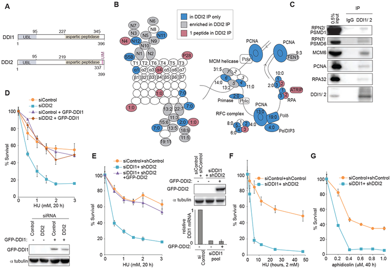
Figure 1: Proteasome shuttles DDI1 and DDI2 function in replication stress response.

(A) Schematic of human DDI1 and DDI2 proteins highlighting their domain structure. Yeast Ddil contains both the UBL and UBA domains typical of shuttle proteins. Human DDI1/2 lack the UBA domain but feature an atypical UBL domain that can bind both ubiquitin and proteasomal ubiquitin receptors. A C-terminal ubiquitin interacting motif (UIM) is specific to human DDI2 (Nowicka et al., 2015; Siva et al., 2016; Trempe et al., 2016). A retroviral aspartyl protease (RVP) domain is also unique to DDI1/2 shuttle proteins (Sirkis et al., 2006). (B) Schematic of the proteasome (RPN, RPT, α, and β subunits) and DNA replication proteins identified to interact with DDI2 after crosslinking with DTSSP. Labeled in blue are proteins identified in the pulldown of GFP-tagged DDI2 but not GFP-only control. In grey are proteins enriched at least twofold in the GFP-DDI2 pulldown compared to GFP control IP. In purple are proteins identified with only one peptide in the GFP-DDI2 pulldown. Ratios in the figure indicate the number of peptides identified in the GFP-DDI2 pulldown to number of peptides in the GFP only control pulldown. (C) Validation of a subset of identified interactions by western blot following IP of endogenous DDI1/2 from cell lysates in the presence of the protein crosslinker DTSSP. All pulldowns for western blot were done in the presence of benzonase. (D) Graphs showing survival of U2OS cells transiently transfected with a pool of DDI2 siRNAs and cross-complemented by stable expression of GFP-DDI1 construct, and a western blot showing GFP-DDI1 expression in these cells. Cells were treated with the indicated doses of HU for 20 h, washed, released, allowed to grow for 7 days, and counted. (E) Graphs showing survival of U2OS cells transiently transfected with a pool of siRNAs against DDI1 or control and stably expressing shDDI2 #1 or control shRNA, that have been complemented with GFP-tagged DDI2, a western blot showing levels of GFP-DDI2, and a graph of relative DDI1 mRNA. Cells were treated as in D. (F) Graphs showing survival of U2OS cells transiently transfected with a pool of siRNAs against DDI1 and stably expressing shDDI2 #1. Cells were treated with 2 mM HU for the indicated time, washed, released, allowed to grow for 7 days, and counted. (G) Graphs showing survival of U2OS cells transiently transfected with a pool of siRNAs against DDI1 or control and stably expressing shDDI2 #1 or control shRNA. Cells were treated with the indicated dose of aphidicolin for 40 h, washed, released, allowed to grow for 7 days, and counted. Error bars represent SEM for 3 replicates. See also Figure S1
