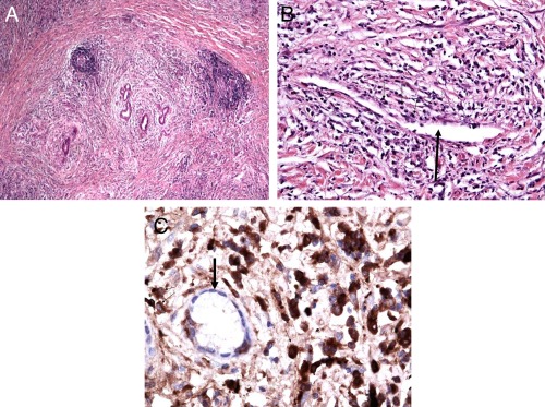
Figure 2.

Histological features in IgG4‐SC. Bile duct resection in IgG4‐SC. (A) Lymphoplasmacytic infiltrate with periductal distribution and a storiform pattern of fibrosis (original magnification ×10). (B) Obliterative phlebitis: a thin‐walled vessel (thick black arrow) surrounded by numerous plasma cells and lymphocytes (thin black arrows) that appear to partially compress the luminal diameter (original magnification ×20). (C) IgG4+ plasma cell infiltration: immunohistochemical staining of the bile duct (thick black arrow) surrounded by abundant IgG4+ plasma cells (thin black arrows; dark brown cells), greater than 50 per high‐power field (original magnification ×40).
