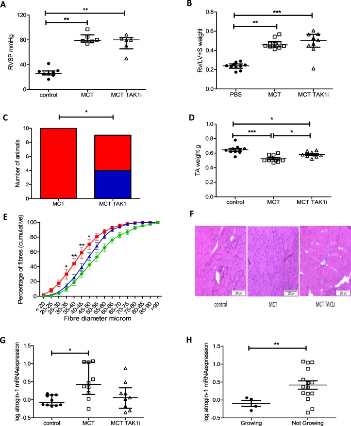
Figure 7.
Transforming growth factor β-activated kinase 1 (TAK1) inhibition prevents tibialis anterior (TA) muscle atrophy in the monocrotaline (MCT) rat as well as preventing weight loss in some animals. (A) Right ventricular systolic pressure (RVSP) in control (● n=8), MCT (□ n=6) and MCT TAK1i (∆ n=6) treated rats (Kruskal-Wallis with Dunn’s correction, p=0.001). (B) Right ventricle/left ventricle plus septal (RV/LV+S) weight in control (● n=10), MCT (□ n=10) and MCT TAK1i (∆ n=9) treated rats (Kruskal-Wallis with Dunn’s correction, p<0.001). (C) Number of MCT animals growing and not growing at the end of the experiment in the MCT (n=10) and MCT TAK1i (n=9) groups (Fisher’s exact test, p=0.033). (D) TA weight in control (● n=10), MCT (□ n=10) and MCT TAK1i (∆ n=9) treated rats (one-way analysis of variance (ANOVA) with Bonferroni correction, p<0.001). (E) Fibre profiles of control ( n=10), MCT (
 n=10) and MCT TAK1i (
 n=9) plotted as mean and SEM of proportion of fibres below the indicated fibre diameter in 5 µm increments (two-way ANOVA, p<0.001 for row, column and interaction, * and ** represent a significant difference between MCT and MCT TAK1 proportions at each 5 µm fibre intervals by Bonferroni). (F) Representative bright-field image of rat TA muscle tissue stained with H&E from which median fibre diameter was determined. (G) Log atrogin-1 mRNA expression in the TA of control (● n=10), MCT (□ n=10) and MCT TAK1i (∆ n=9) treated rats (Kruskal-Wallis with Dunn’s correction, p=0.018). (H) Log atrogin-1 mRNA expression in the TA of MCT-treated rats growing (● n=4) and not growing (□ n=15) at the end of the experiment (Mann-Whitney U test, p=0.032).

