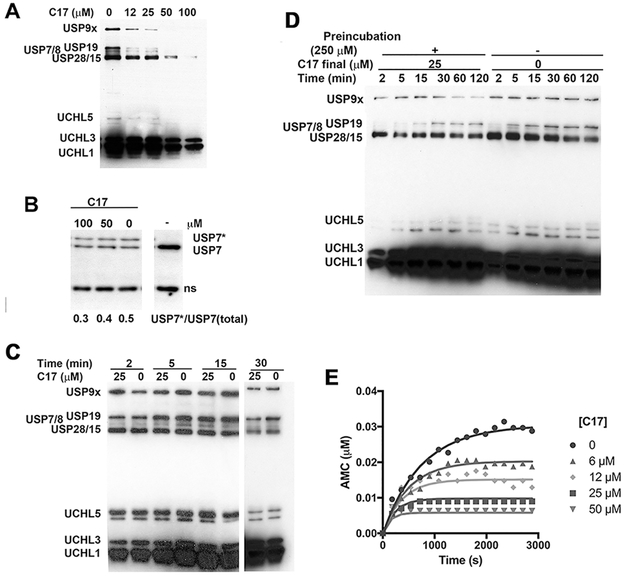
Figure 3. Characterization of DUB inhibition by C17.
A. A lysate of HEK 293T cells (1.5 mg/ml) was treated with C17 (25 μM) or DMSO followed by HA-Ub-VS (1.5 μM). Aliquots were removed and analyzed for HA. B. A HEK 293T cell lysate was incubated with C17, treated with HA-UbVS, analyze by SDS-PAGE and immunoblotted with anti-USP7. USP7* denotes USP7 modified by HA-Ub-VS. An intervening lane was removed for clarity. C. A lysate of HEK 293T cells (1.5 mg/ml) was treated with C17 (25 μM) or DMSO simultaneously with HA-Ub-VS (1.5 μM). Aliquots were removed and analyzed for HA. Intervening lane removed for clarity. D. A lysate of HEK 293T cells (15 mg/ml) was treated with either C17 (250 μM) or DMSO for 30 min. After this time, lysate was diluted tenfold and HA-Ub-VS (1.5 μM) was added. Aliquots were removed and analyzed by HA blot. E. Representative progress curve. Purified recombinant His6USP9x (0.7 nM) was mixed with Ub-AMC (300 nM) and C17 in 1% DMSO and reaction was monitored by measuring AMC release on a 96 well plate reader. Lines denote fits to a single step inactivation mechanism as described in Material and Methods.

