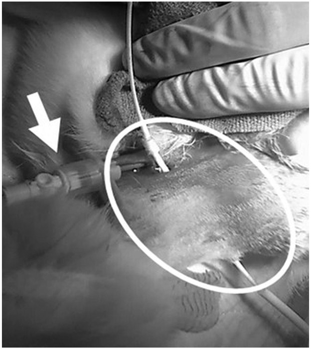
Figure 1.

Intraoperative photograph of a female New Zealand White rabbit after insertion of the peel-away sheath (arrow) into the right internal jugular vein and interscapular subcutaneous tunneling of the 4.2-F catheter from the venotomy to the skin exit site (circle).
