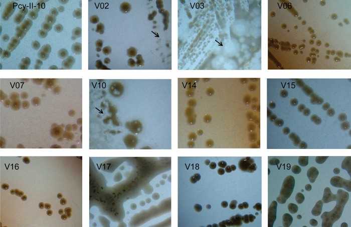
Fig 1. Colony morphology of PcyII-10 and phage-resistant variants.
Colonies formed by PcyII-10 and eleven variants plated at P3 on LB agar were photographed after 24 h at 37°C. The arrows point to lysis zones caused by phages. Variant V03 produces small colonies with irregular shapes. Variants V06, V07, V14 and V16 produce a reddish-brown pigment, the pyomelanin. Variants V17 and V19 were mucoid.

