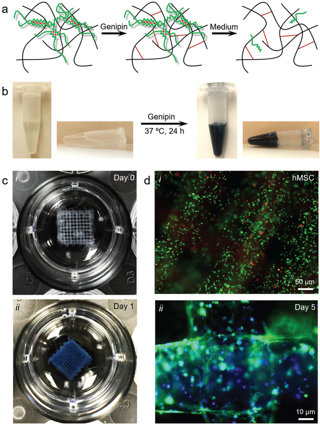
Figure 3.
Templated microfluidic bioprinting of the alginate–gelatin bioink. a) Schematics showing the dual-step crosslinking process and subsequent removal of alginate. b) Photographs showing the crosslinking process of gelatin by genipin. c) Photographs showing a bioprinted five-layer construct at c-i) day 0 and c-ii) day 1 post-bioprinting. d) Fluorescence microscopic images showing d-i) live/dead staining (live cells in green and dead in red) at day 0, as well as d-ii) F-actin/nuclei staining (F-actin in green and nuclei in blue) at day 5 of the encapsulated hMSCs in the bioprinted constructs.

