Fig. 6.
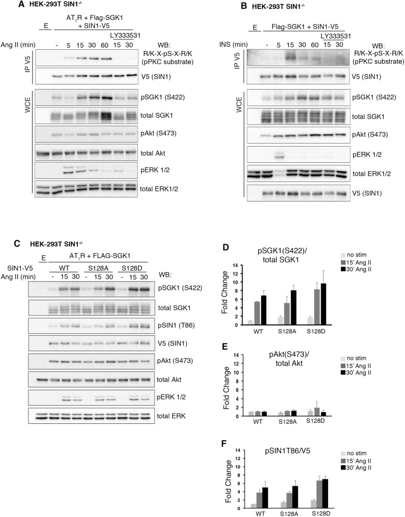
AngII and insulin signaling trigger phosphorylation of SIN1 at S128 but SIN1 S128 phosphorylation is not required for SGK1 S422 phosphorylation. (A) Western blot (WB) analysis of whole-cell extracts (WCEs) and anti-V5 immunoprecipitations (IPs) derived from SIN1−/− cells transfected with Flag–SGK1, SIN1–V5 and AT1R. Cells were serum starved overnight before treating with AngII (200 nM) for the times shown. Where indicated, the cPKC inhibitor LY333531 (200 nM), was added. E, empty vector. (B) WB analysis of WCEs and anti-V5 IPs derived from SIN1−/− cells transfected with Flag–SGK1, SIN1–V5 and empty vector. Cells were serum starved overnight before treating with insulin (INS) (200 nM) for the times shown. Where indicated, the cPKC inhibitor LY333531 was added. Results are in A and B show a representative of n=2 biological replicates. (C) WB analysis of WCEs derived from SIN1−/− cells transfected with AT1R, Flag–SGK1 and the indicated SIN1–V5 constructs. Cells were serum starved overnight and then treated with AngII (200 nM) for the times shown. Results are representative of n=3 biological replicates. (D–F) Quantification of pSGK1 S422, pAkt S473 and pSIN1 T86 levels from WBs as shown in C, corrected for total protein level; values represent fold change from unstimulated ‘WT SIN1’. Data are represented as mean±s.e.m. (n=3 biological replicates).

