Fig. 7.
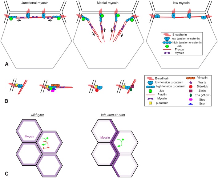
Models for Jub localization and action at AJs. (A) Illustration of the hypothesis that formation of the high-tension conformation of α-catenin that binds Jub depends upon both the amount of tension (arrows) in actomyosin filaments (red lines) and its orientation relative to AJs. Jub binding requires tension applied perpendicular to the membrane. For simplicity, AJs are only shown along one side of the cell (black lines), not on other sides (gray lines). (B) Illustration of four different types of AJ complexes identified in wing imaginal disc cells at different locations around the cell circumference. (C) Illustration of how myosin distribution is altered in the absence of jub, step or sstn. Tension-dependent recruitment of myosin creates a positive-feedback loop, which, in the absence of the negative-feedback loop provided by Jub, Sstn and Step, we hypothesize results in accumulation of myosin in multicellular cables and depletion from other cell–cell junctions.

