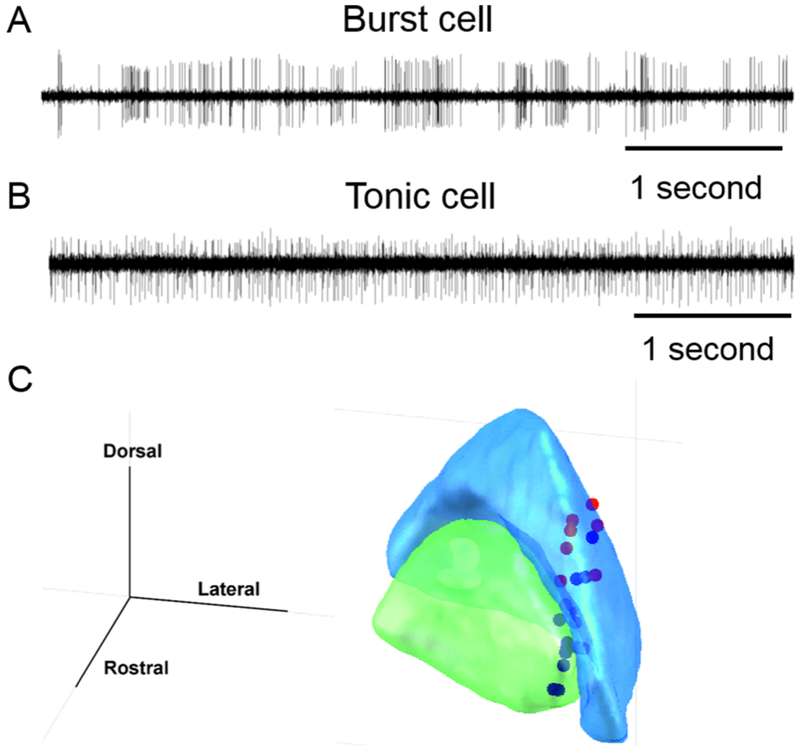
Fig. 2:

(A) Example of single unit recording from the burst neuron in globuls pallidum interna (GPi). Amplitude of electrical activity (mV) is plotted on y-axis, while x-axis depicts time. The time series illustrates bursts of spikes in action potential with intervening pause in the activity. Such activity pattern is typical for the pallidal burst neurons. (B) Example of single unit activity recorded from the tonic neuron in globus pallidum internus. In this example, in contrast to the burst neuron, the spikes in action potential are nearly continuous with no epochs of silence. The response is typical of pallidal tonic neurons. (C) Locations of the head movement sensitive pallidal neurons overlaid on the 3D outline of GPe (blue) and GPi (green).
