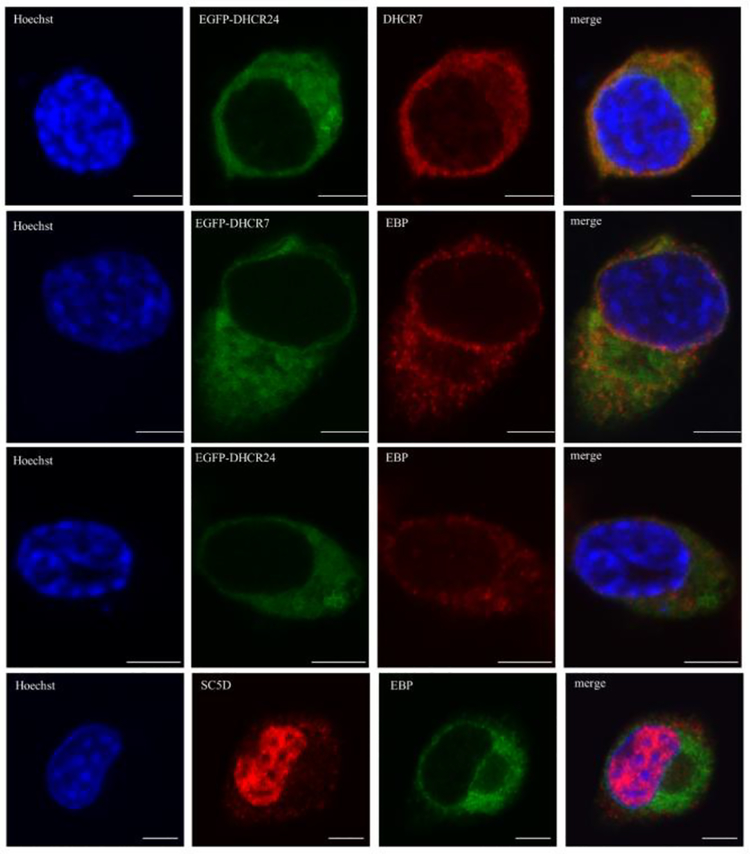
Figure 5. DHCR7, DHCR24, EBP and SC5D cholesterol biosynthesis enzymes show only a partial co-localization.
Neuro2a cells were transiently transfected with EGFP-DHCR7 and EGFP-DHCR24 fusion constructs (rows 1–3) and labeled with anti-EBP and anti-DHCR7. Control Neuro2a cells were double stained with anti-SC5D and anti-EBP antibodies (bottom row). Cells were analyzed by confocal microscopy. Nuclei were visualized by Hoechst dye. Original images were collected with a 60× oil objective. Calibration bar, 5 μm. Note that DHCR7 and DHCR24 showed the most prominent co-localization throughout the cellular compartments, while the more proximal sterol biosynthesis enzyme SC5D in addition to overlap with DHCR7, DHCR24 and EBP within ER is also present in the nucleus.

