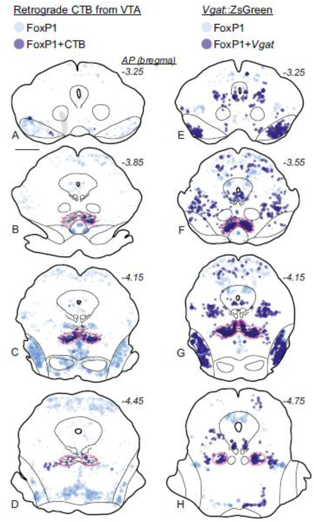
Fig. 11.
FoxP1 neurons in the RMTg project to VTA and express Vgat in mice. a-d. FoxP1 is found throughout the midbrain, but is particularly dense in the RMTg (magenta dashed lines). Within the midbrain, neurons double-labeled with FoxP1 and retrograde tracer (CTB) from VTA are almost exclusively found in the RMTg. Translucent gray shows location where heavy CTB fiber expression precluded accurate neuronal labeling. e-h. FoxP1 neurons express Vgat in several regions in transgenic Vgat::ZsGreen mice, including the RMTg (f), SNr (e), and NLL (g). Notably, despite the large numbers of Vgat neurons in the VTA, very few double labels with FoxP1 are visible (e). Scalebar = 1 mm.

