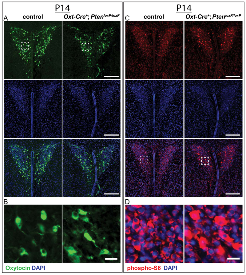
Figure 7.
Assessment of Oxt immunoreactive cells and phospho-S6 enrichment in the PVN at P14. (A,B) Representative images of P14 control and Oxt-Cre+; PtenloxP/loxP PVN (A) immunostained with anti-Oxt (green) and DAPI (blue), with enlargements showing cellular resolution (white square, B). (C,D) Representative images of P14 control and Oxt-Cre+; PtenloxP/loxP PVN (C) immunostained with anti-phospho-S6 (red) and DAPI (blue), with enlargements showing cellular resolution (white square, D). Scale bars: 200 μm (A,C), 20μm (B,D).

