
Figure 2.
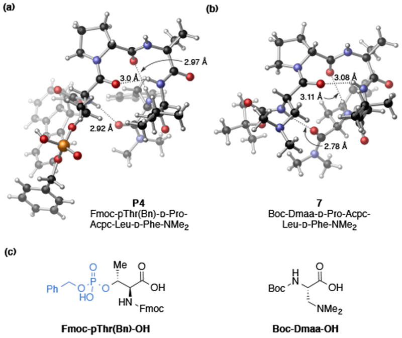
Comparison of X-ray crystal structures of pThr- and Dmaa-embedded peptides. (a) X-ray structure of P4. Two 1,4-dioxane solvent molecules omitted for clarity (see SI for details). (b) X-ray structure of 7. Two distinct packing polymorphs were observed in the unit cell (7a, 7b), only one is shown for clarity (See SI for details). (c) Catalytically active residues (pThr and Dmaa) embedded in i position of conserved peptide sequence.