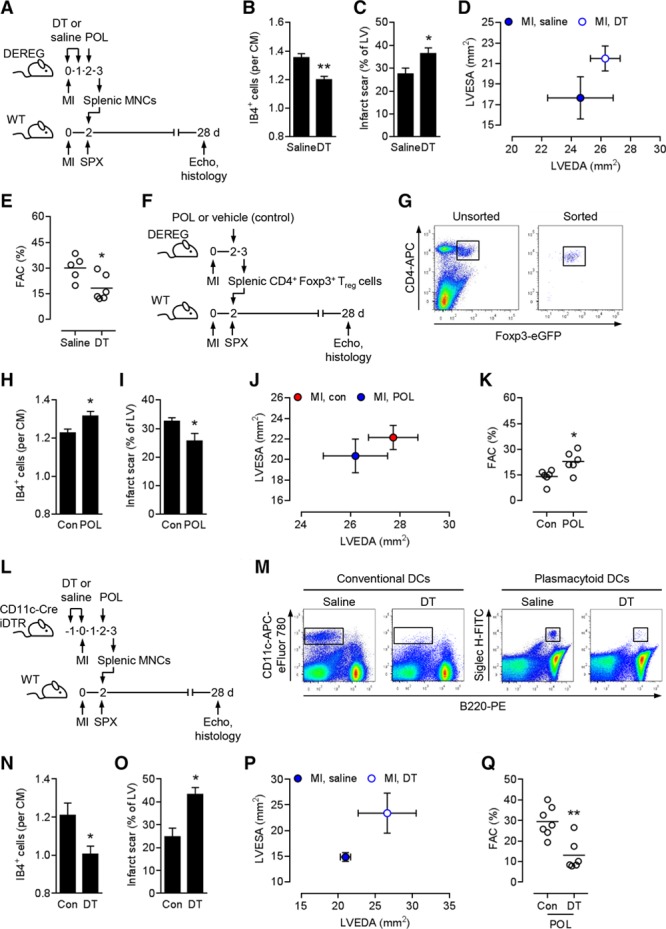
Figure 5.

Splenic regulatory T cells are required and sufficient for the therapeutic effects of POL5551. A, Experimental setup for B through E. Myocardial infarction (MI) was induced in DEREG donor and wild-type (WT) recipient mice. Donor mice were intraperitoneally injected with diphtheria toxin (DT) or saline immediately before and 24 hours after MI. Two days after MI, donor mice were intraperitoneally injected with POL5551 (POL), and splenic mononuclear cells (MNCs) were isolated 24 hours later. Two days after MI, recipient mice were splenectomized (SPX) and then infused intravenously with donor MNCs. Echo indicates echocardiography. B through E, Five mice treated with MNCs from saline-injected donors, 6 mice treated with MNCs from DT-injected donors. B, Fluorescein-labeled isolectin B4 (IB4)+ capillary density in the infarct border zone 28 days after MI. CM indicates cardiomyocyte. C, Scar size 28 days after MI. LV indicates left ventricle. D, LV end-diastolic area (LVEDA) and LV end-systolic area (LVESA) as determined by echocardiography 28 days after MI. E, Fractional area change (FAC). F, Experimental setup for H through K. MI was induced in DEREG donor and WT recipient mice. Two days after MI, donor mice were intraperitoneally injected with POL5551 or vehicle only (Con), and splenic CD4+ Foxp3+/eGFP+ regulatory T (Treg) cells were isolated 24 hours later. Two days after MI, recipient mice were splenectomized and then infused intravenously with donor Treg cells. G, Representative flow cytometry panels showing unsorted splenic MNCs (left) and sorted splenic Treg cells (right) that were used for transplantation. CD4+ Foxp3+/eGFP+ Treg cells are highlighted in both panels. H through K, 6 mice that received Treg cells from vehicle-only–treated donors, 6 mice that received Treg cells from POL5551-treated donors. H, Capillary density in the border zone 28 days after MI. I, Scar size 28 days after MI. J, LVEDA and LVESA 28 days after MI. K, FAC. L, Experimental setup for N through Q. MI was induced in CD11c-Cre iDTR donor and WT recipient mice. Donor mice were intraperitoneally injected with DT or saline 24 hours before and immediately before MI. Two days after MI, donor mice were intraperitoneally injected with POL5551, and splenic MNCs were isolated 24 hours later. Two days after MI, recipient mice were splenectomized and then infused intravenously with donor MNCs. M, Representative flow cytometry panels showing non–dendritic cell (DC)–depleted (saline) and DC-depleted (DT) splenic MNCs that were used for transplantation. DCs are highlighted in all panels. N through Q, Seven mice treated with MNCs from saline-injected donors, 6 mice treated with MNCs from DT-injected donors. N, Capillary density in the border zone 28 days after MI. O, Scar size 28 days after MI. P, LVEDA and LVESA 28 days after MI. LVESA: P<0.05 (2-independent-sample t test). Q, FAC. *P<0.05, **P<0.01 (2-independent-sample t tests).
