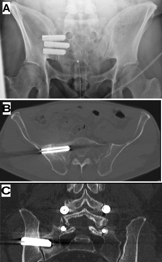
Fig. 7.
Imaging of typical configuration of implants. Fig. 7-A Inlet-view pelvic radiograph. Fig. 7-B A 12-month CT image from a different subject showing no radiolucencies around the first implant. Fig. 7-C A 12-month CT image from another subject showing radiolucency around the second implant in the sacrum.

