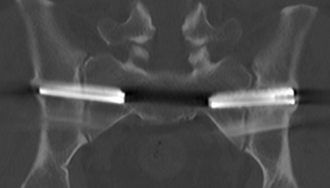
Fig. 8.
A 12-month CT image depicting bilateral implants with bone apposition along the entire length of the superior and inferior sides of both implants. Also, there is bone overgrowth at the outer iliac cortex (the left side is greater than the right side), suggesting complete implant integration into the ilium. However, there is little bone apposition along the implants within the joint.

