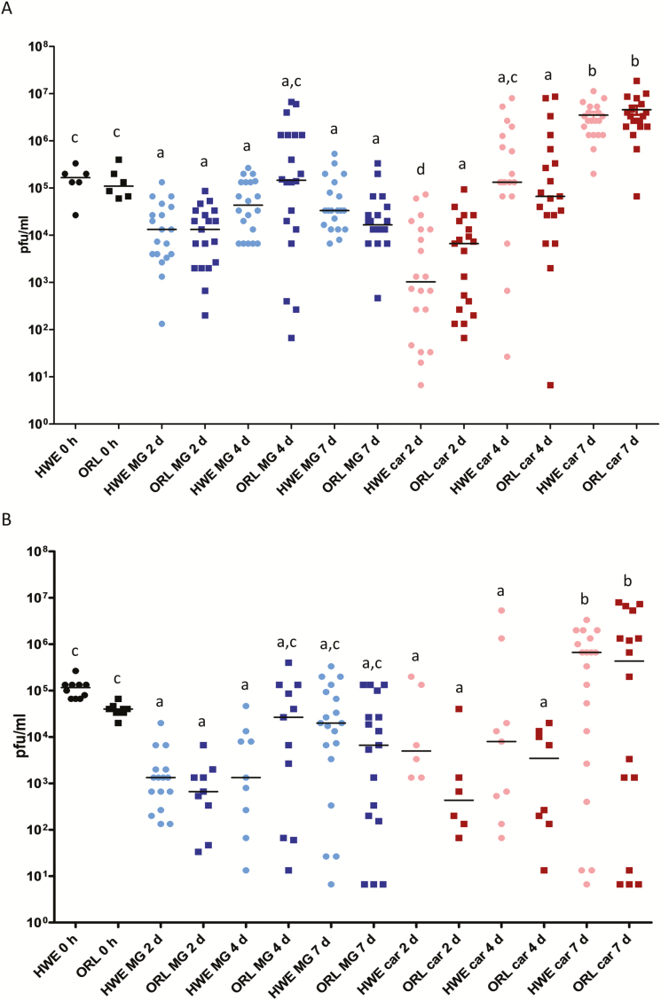
Fig. 3.
Intensities of MAYV IQT and TRVL infections in midguts and carcasses of Ae. aegypti HWE and ORL. (A) MAYV IQT (artificial bloodmeal titer: 2.0 × 10⁶ plaque forming units (PFU)/ml) and (B) MAYV TRVL (artificial bloodmeal titer: 5.0 × 10⁶ PFU/ml) titers in midguts (n = 20) and carcasses (n = 20) of individual HWE and ORL females analyzed at 2, 4, and 7 d pibm by plaque assays in Vero cells. Each data point represents the MAYV titer of an individual midgut or carcass. For 0 h, only whole-body females were assayed. Only infected mosquitoes were included in the analysis. Black bars indicate medians. Different letters indicate median titers that were significantly different from each other (Mann–Whitney U test). Significantly different comparisons: P values ranged from < 0.0001 to 0.0016 in (A) and from < 0.0001 to 0.0004 in (B).

