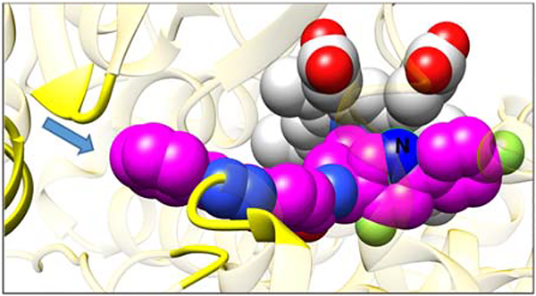
Figure 5. Expected (VFV-based) position of the 1-(pyridin-3-yl) nitrogen atom (marked as N) of compound 1 in the CYP51 structure.

The carbon atoms of the heme and the inhibitor are light gray and magenta, respectively. Oxygens are red, nitrogens are blue, chlorines and fluorines are green. The arrow shows entrance into the enzyme access channel. The protein ribbon around the entrance is yellow, the rest of the ribbon is semitransparent gold.
