
Figure 2: Three crystallographic sites for cholesterol on three different GPCRs.
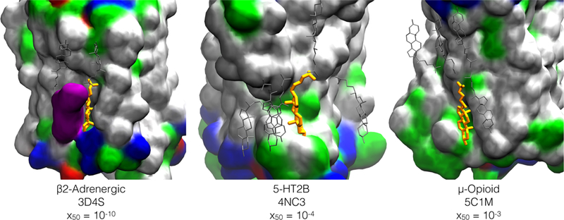
Protein is drawn in space-filling and colored by residue type : hydrophobic (white), polar (green), acidic (red), basic (blue). A second crystallographic cholesterol in the β−2adrenergic structure 3D4S is also shown in space-filling, colored purple. A subset of the additional cholesterol molecules placed by CHARMM-GUI Membrane Builder for a mixture of 7:3 POPC:CHOL are in silver. Crystallographic cholesterol molecule in sites characterized in this work are in orange, and are residue 402,1203, and 404 in structure 3D4S, 4NC3, and 5C1M respectively. The corresponding half-saturation cholesterol fraction x50 is from Table 6; for the µ-opioid receptor, pocc < 50% for the entire cholesterol range.