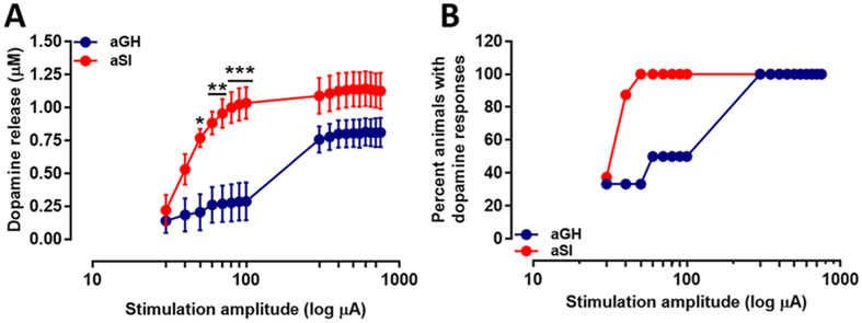
Figure 4.
Dopamine release elicited in NAc core by low and high stimulus intensities. (A) Dopamine release measured following single pulse electrical stimulation of low and high intensities. Dopamine release in aSI rats was measured to be significantly greater compared to dopamine release in aGH rats. Bonferroni posthoc analysis revealed a significant potentiation of dopamine release at the 50, 60, 70, 80, 90, and 100 μA stimulation intensities. (B) Percentage of slices (animals) in aGH and aSI groups in which dopamine was evoked at low and high stimulus intensities. Group housed, aGH, blue, n = 6; Socially isolated, aSI, red, n = 8; *p < 0.05; **p < 0.01; ***p < 0.001.

