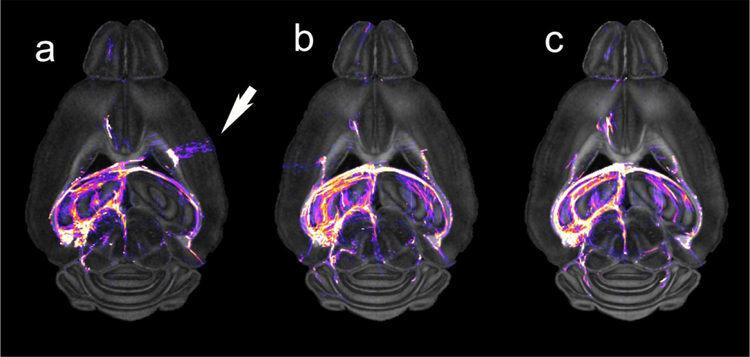
Figure 4.
Dorsal NQA image from a C57BL/6J mouse acquired with a) 16 angles; b) 46 angles; c) 120 angles demonstrate the value of increased angular sampling. Tractography was generated by seeding the left hippocampus. The white arrow highlights a false positive tract in the 16-angle data that does not appear in the 46 and 120 angle data.

