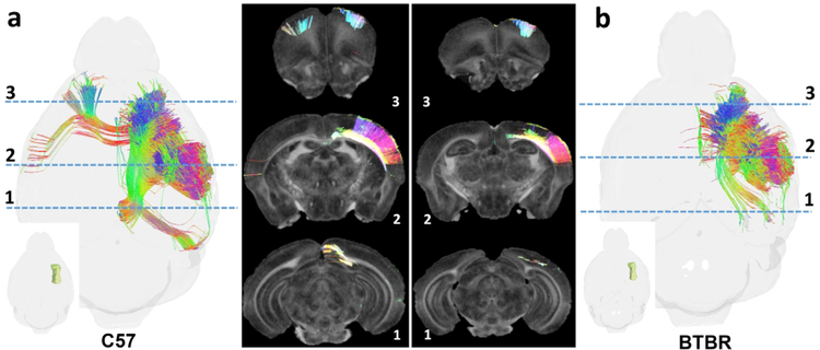
Figure 8.
Comparison of tractography within the forelimb representation of the right primary somatosensory cortex in two murine strains (individual specimens): C57BL/J6 (a), a common mouse model in biomedical research, and BTBR (b) a murine model that bears phenotypic resemblance to human autism spectrum disorders (Meyza and Blanchard, 2017, Scattoni et al., 2008). Images to the far left and right in (a) and (b) show tractography as seen from the dorsal aspect of the mouse brain; Insets localize the forelimb representations. Dashed lines identify the locations of coronal NQA images from the same datasets with in-plane streamlines superimposed. Note strain-specific differences in commissural connections and longitudinal associational connections in the ipsilateral hemispheres.

