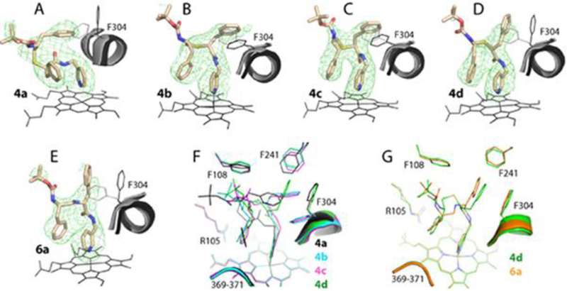
Figure 4.

Crystal structures of CYP3A4 bound to compounds from the methyl-pyridyl subseries. A-E, The binding mode of 4a-d and 6a, respectively. The adjacent I-helix and Phe304 in the inhibitory complexes and in water-bound CYP3A4 (4I3Q structure) are shown in black and gray, respectively. Polder omit maps contoured at 4σ level are shown as green mesh. F and G, Structural overlays of 4a-d and the 4d-6a pair, respectively.
