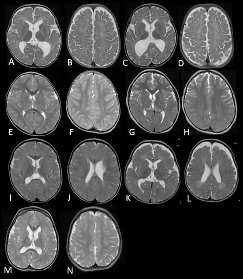
Figure 1: Evidence of CNS myelin defects in affected individuals carrying FIG4 variants. MRI images.

Axial T2 weighted MR images of patient 1(family 1) at 10 months (A,B) and 27 months (C,D) demonstrating complete absence of myelination in cerebral white matter and internal capsule with no improvement on follow up. Patient 2 (family 2) at 30 months (E,F) and 8 years (G,H) demonstrates high signal in the posterior limb of the internal capsule and diffuse high signal in the posterior periventricular and deep cerebral white matter. Family 3, sibling 1 at 25 months (I,J) demonstrates mild high signal in cerebral white matter with myelination present in deep and subcortical white matter. Family 3, sibling 2 at 18 months (K,L) demonstrates more striking hypomyelination with very little normal myelin visible. The MRI of patient 5 in family 4 at 3 years of age was largely normal with non-specific high signal in the periventricular and deep parietal white matter (M,N).
