
Figure 5. Patient fibroblasts from Family 1 and Family 2 exhibit the characteristic vacuolization caused by deleterious variants of FIG4.
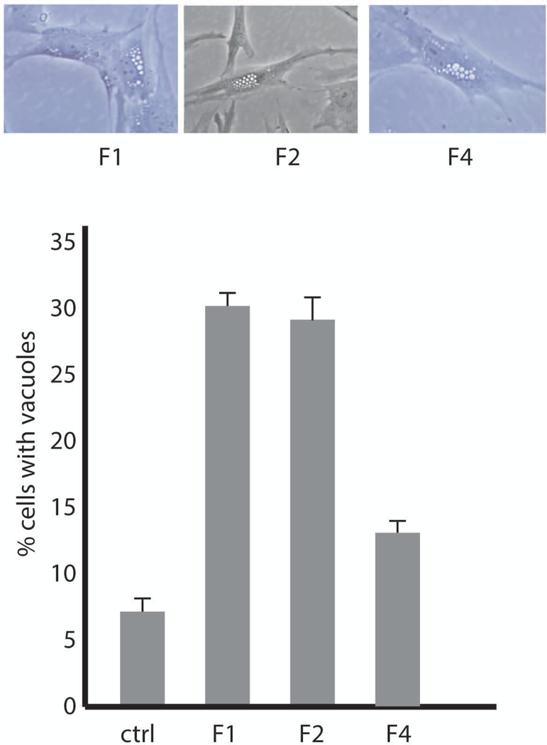
A) Live cell microscopy of cultured fibroblasts from affected individuals. B) Quantitation of the extent of vacuolization. Values represent means ± SD.