Figure 3.
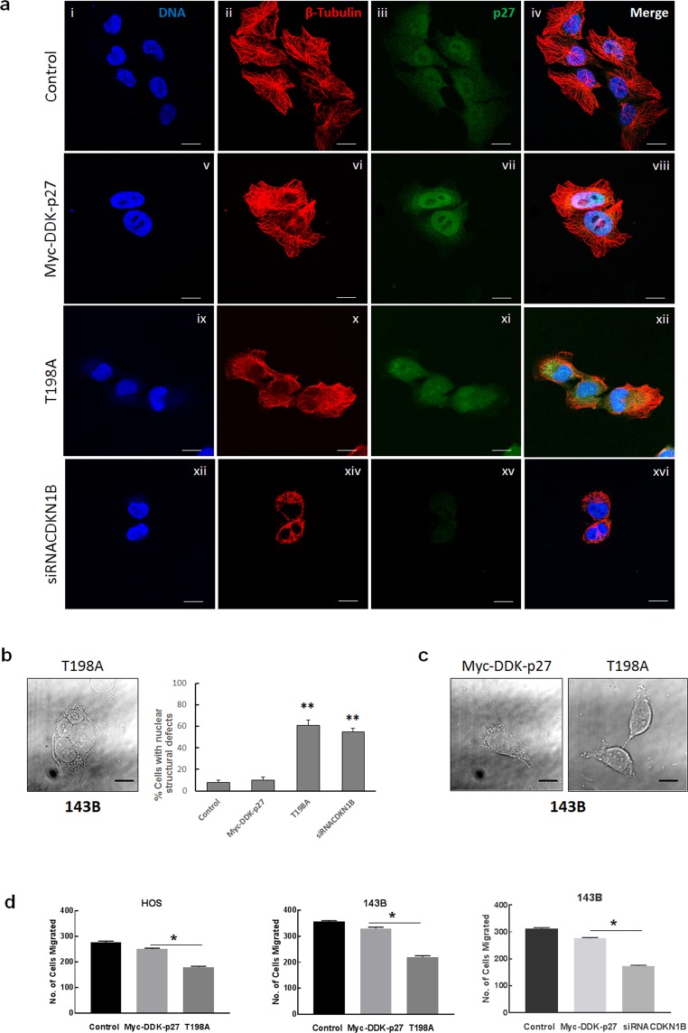
p27 and STMN1 interaction regulates microtubule stabilization and migration of osteosarcoma cells. (a) 143B untransfected controls and cells expressing recombinant wild-type p27 (Myc-DDK-p27), T198A mutant p27 (T198A) and p27 depletion were subjected to immunofluorescence staining with antibodies against β-tubulin and p27. β-tubulin is colored red; p27 is colored green; nuclei were stained with Hoechst dye and are colored blue. Magnification is 63x; scale bars = 25 μm. (b) 143B cells were imaged using differential interference contrast (DIC) microscopy and analyzed for nuclear structural defects. Left panel: Representative image of a polynucleated cell expressing T198A mutant p27. Right plot: Each column represents percentage of nuclear structural defects for 100 cells for indicated experimental groups. *p < 0.5. (c) DIC image of 143B control cell and apoptotic cell expressing T198A mutant p27. Magnification is 63x; scale bars = 25 μm. (d) The matrigel invasion assay was conducted using HOS (left plot) and 143B cells (middle and right plots) expressing recombinant wild-type p27, T198A mutant p27 and reduced p27 (siRNACDKN1B). Data is shown as mean + SE from three experiments; *represents p < 0.05.

