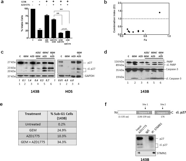
Figure 4.
AZD1775 causes accumulation of p27 form in osteosarcoma cells. (a) 143B cells were incubated with gemcitabine (GEM), AZD1775 or the indicated combinations of both agents and cell viability was measured using the cell titer blue assay. Data is shown as mean + SE; *represents p < 0.05; n = 6. (b) Chou-Talalay analysis of synergy in 143B cell lines for GEM/AZD1775 treatment. CI < 1 indicates synergy of agents. Drug dosing concentrations are listed in Supplementary Table S1. (c) Immunoblot analysis of lysates extracted from 143B cells treated with GEM and AZD1775 as indicated using anti- p27 antibody. GAPDH was the protein loading control. Relative expression of cleaved p27 (23 kDa) was quantified from immunoblots using Image J. Results shown are mean + SE from three experiments. (d) Immunoblot analysis of the apoptotic markers cleaved PARP and cleaved caspase-3 were examined by immunoblot analysis using cell lysates isolated from cells exposed to the indicated treatments with GEM and AZD1775. (e) The cell cycle distribution was analyzed by flow cytometry using propidium iodide DNA staining following the indicated treatments of cells with GEM and AZD1775. The percent of sub-G1 cells is listed. The results are representative of three independent experiments. (f) HOS cells treated with AZD1775 were lysed and subjected to immunoprecipitation with anti-STMN1 antibody; p27 was detected by immunoblot with anti-p27 antibody (top panel) and STMN1 was detected with anti-STMN1 antibody (lower panel).

