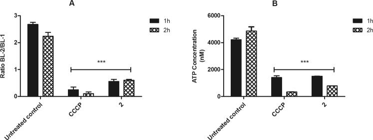
Figure 3.
Evaluation of mitochondrial membrane potential in L. (L.) infantum promastigotes treated with compound 2 (190 μM). (A) JC-1 dye fluorescence was measured by flow cytometry (excitation 488 nm and emission 530/574 nm) after 1 and 2 h of incubation. Untreated promastigotes and treated with CCCP (100 μM) were used to achieve minimal and maximal depolarization, respectively. Fluorescence is reported as the ratio between the emission channels BL2/BL1. (B) Evaluation of ATP concentration. ATP was measure spectrofluorimetrically after 1 and 2 h of incubation. Untreated promastigotes and treated with CCCP (25 μM) were employ to achieve minimal and maximal depolarization, respectively. Results are expressed in nM. A representative experiment is shown. ***p < 0.0001.

