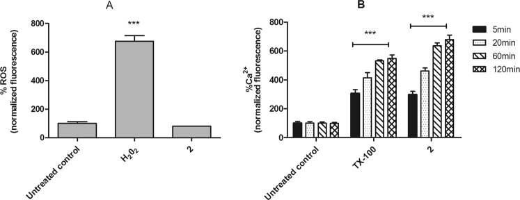
Figure 4.
Evaluation of reactive oxygen species (ROS) and intracellular calcium levels in L. (L.) infantum promastigotes treated with compound 2 (190 μM). (A) H2DCFDA dye fluorescence was measured by spectrofluorimetrically (excitation 485 nm and emission 520 nm) after 2 h of incubation. Untreated promastigotes and treated with H2O2 (400 μM) were used to achieve minimal and maximal depolarization, respectively. (B) Fura-2 AM dye fluorescence was measured spectrofluorimetrically (excitation 360 nm and emission 500 nm) after 5, 20, 60 and 120 min of incubation. Untreated promastigotes and treated with TX-100 (0.5%) were used to achieve minimal and maximal depolarization, respectively. Fluorescence is reported as percentage relative to untreated promastigotes (100%). A representative experiment is shown. ***p < 0.0001.

