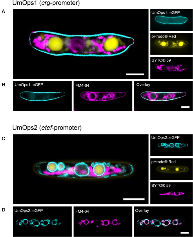
FIGURE 5.

Life-cell fluorescence microscopic analysis of the localization of eGFP-tagged UmOps1 (A,B) and UmOps2 (C,D) after heterologous expression in U. maydis sporidia. Images were obtained with a 3D-SIM. Sporidia were either co-stained for mitochondria (SYTO®59) and vacuoles (pHrodo®Red; A,C), or for vacuolar membranes (FM4-64; B,D). Expression of UmOps1-eGFP was driven by the arabinose-inducible crg-promoter, expression of UmOps2-eGFP by the constitutive etef-promoter. Localization of UmOps2-eGFP after arabinose-induced expression is shown in Supplementary Figure S8. Note that UmOps1 is mainly located in the plasma membrane whereas UmOps2 is present in the vacuolar membranes. Scale bars represent 3 μm.
