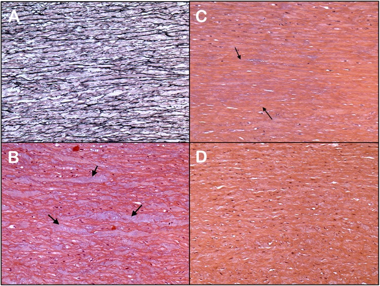
FIGURE 1.
Different histological features observed in aortas of BAV patients (A–C) and the normal aorta (D): (A) Fragmentation and loss of the elastin fibers (limited to 1 or 2 lamellar units; mild grade) (Elastic stain; Original X10). (B) Inter-lamellar degeneration with mucoid replacement (MEMA) (H&E stain; Original X10). (C) Moderate loss of nuclei of vascular smooth muscle cells (involving 4 to 10 lamellar units; area between arrows) (H&E stain; Original X10). (D) Normal ascending thoracic aorta. The media shows no mucoid accumulation in the extracellular matrix or loss of smooth muscle cells (H&E stain; Original X10).

