
FIGURE 4.
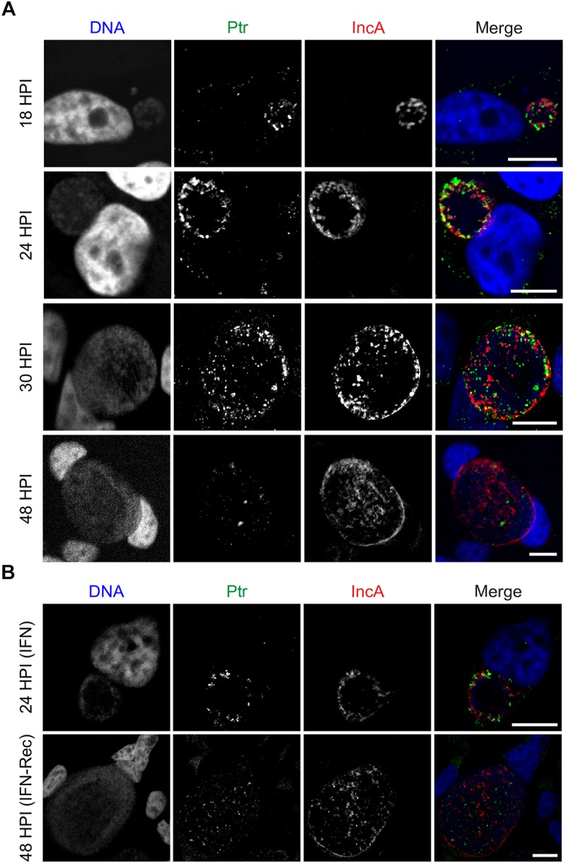
Ptr is expressed at mid to late stages of C. trachomatis developmental cycle. (A) Representative micrographs of HeLa cells infected with LGV-L2 at 18-24-30-48 hpi. (B) Representative images of HeLa cells infected with LGV-L2 during IFNγ treatment (IFN: cells were pre-treated for 24 h with 15 ng/mL of IFNγ, then infected for 24 h in presence of IFNγ) and upon recovery from IFNγ-induced stress (IFN-Rec: at 48 hpi, 24 h after removal of IFNγ and addition of tryptophan). Anti-Ptr (green) shows Ptr expression and anti-IncA (red) marks the boundaries of the inclusion membrane. Confocal images are depicted. Scale bars, 10 μm.